Credit: Wilhelm Röntgen
X-rays are common today, but their accidental discovery 125 years ago was celebrated around the world as a scientific miracle and earned Wilhelm Röntgen the first Nobel Prize in physics.
Röntgen was working in his lab in Germany, trying to replicate the experiments of other scientists with an electrified cathode tube, when he noticed some barium-painted cardboard nearby fluorescing.
Puzzled, he turned off the lights, but it continued to glow. He realized the cathode tube was emitting something other than light. When he went to move the barium board, he noticed a lead plate cast a shadow on it.
He began to try to create images, placing objects between the tube and the cardboard. He realized the mysterious rays were penetrating soft objects but not hard, and was shocked to see, in one image, what looked like the bones of his hand!
Recognizing the potential, he tried to capture a clearer image of bones. He finally produced one of his wife’s hand and published it in an article.
Not knowing what the rays were, he named them after the mathematical unknown, “x.”
Worldwide acclaim was swift. Within a month, he was called in front of Kaiser Wilhelm to demonstrate and awarded a prestigious medal on the spot.
Within a year, doctors in the Balkan war were using X-rays to find bullets and broken bones, and scientists the world over were experimenting with them… without understanding their risks.
But that’s a story for another EarthDate.
Background
Synopsis: 125 years ago, Wilhelm Röntgen discovered he could make the invisible visible by using a new type of electromagnetic radiation that he called “X-rays”—he didn’t know what they were, so he named them after the mathematical variable “x.” Touted as a medical miracle in the early 1900s, today they are extensively used for everything from medical diagnostics, to material analysis, to security screening. They have even been used to determine the composition of Martian soil.
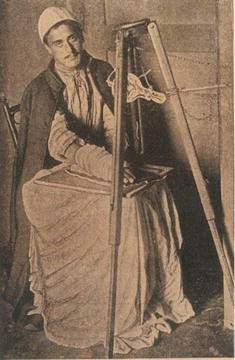
Credit: Esad Feyzi
- Today, most of us think of X-rays as commonplace, but in the early 1900s, they were an amazement for their ability to let people see inside the human body and through solid objects.
- Most of us have had an X-ray at the dentist’s or doctor’s office. Fluoroscopes and CT (computed tomography) scans also use X-rays. Anyone who has taken luggage through an airport has probably had their baggage screened by X-ray machines too.
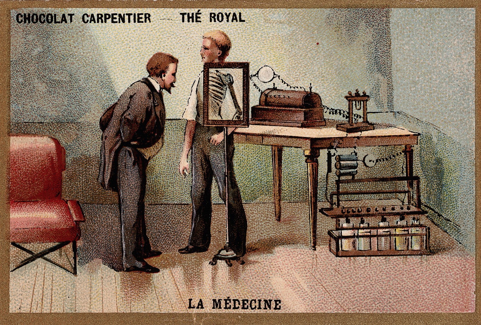
- In 1896, when the first X-ray demonstrations occurred, people were shocked to see bones inside a living body for the first time.
- X-rays revolutionized medicine. They were immediately put to use in medical situations and were a great help to doctors during the Greco-Turkish War of 1897, as they enabled surgeons to view bullets and broken bones in their patients.
- X-rays were accidentally discovered on November 8, 1895, by scientist Wilhelm Röntgen at his laboratory in the Würzburg Physical Institute of the University of Würzburg, Bavaria, Germany.
- Röntgen wasn’t trying to discover X-rays at all. He was setting up an experiment testing cathode rays in a variety of vacuum tubes to assess the external effects of various tube configurations.
- Scientists had discovered that when voltage is applied in a vacuum tube, invisible cathode rays move from a negative cathode toward a positive anode.
- The scientific community eventually figured out that cathode rays were beams of negatively charged particles that they named electrons. These were the first subatomic particles to be identified.
- Röntgen was repeating the experiments of other scientists to confirm their results and better understand the phenomenon.
- During one test, he noticed that a small cardboard screen painted with barium platinocyanide began to fluoresce. He repeated the test and found that even when all the light from the tube was blocked, the fluorescence still occurred.
- In another accident, he found that a piece of lead held between the source and the fluorescent screen cast a shadow. He then realized he was dealing with an invisible form of energy that was similar to light’s electromagnetic energy.
- Röntgen found that by directing cathode rays toward objects placed on photographic plates, he could capture their shapes. He found he had even captured a ghostly image of the bones in his fingers!
- Röntgen wasn’t trying to discover X-rays at all. He was setting up an experiment testing cathode rays in a variety of vacuum tubes to assess the external effects of various tube configurations.
- For the next 6 weeks, he studied the new phenomenon and documented his findings. He published his first paper, titled “On A New Kind of Rays” (Ueber eine neue Art von Strahlen), on December 28, 1895.
- On December 22, 1895, Röntgen took an X-ray of his wife’s left hand, and on January 1, 1896, he presented it to colleagues at the Physik Institut at the University of Freiburg, Germany.
- The news spread quickly. By January 5th, the new type of radiation had been reported in the Weiner Presse, an Austrian newspaper, and soon the story broke around the world.
- On January 13th, Röntgen was summoned to show his discovery to Kaiser Wilhelm II, the emperor of Germany, who awarded him the Prussian Order of the Crown, Second Class on the spot.
- At a lecture on January 23, 1896, Röntgen took an X-ray of a colleague’s left hand to publically demonstrate the ease of using this medical magic.
- X-rays penetrate human flesh but not higher-density bone or metal, which cast a “shadow” on photographic plates exposed to X-radiation. These bright shadows are what we see on our medical X-rays.
- People weren’t just excited about X-rays as a revolutionary medical tool. The technology quickly gained popularity in public exhibitions and lectures. Everyone seemed morbidly curious to see the skeleton of a living person for the first time.
- In 1896 and 1897, Röntgen published two more papers describing medical and metallurgical applications for X-rays.
- In 1901, he received the first ever Nobel Prize in physics for his work on X-rays.
- Since he didn’t know what his discovery was, he temporarily named the rays after the mathematical name for an unknown variable, “x.”
- The name stuck in many countries. However, in his native Germany they are called Röntgen rays (Röntgenstrahlen), and the medical plates, or X-radiograms, are called Röntgenograms.